A lung X-ray (chest radiograph) captures images of the lungs and chest cavity to identify abnormalities like pneumonia, tuberculosis, pleurisy, tumors, and chronic bronchitis. It is one of the first diagnostic tools used when patients report respiratory symptoms.
When It’s Recommended:
- Persistent cough
- Shortness of breath
- Fever or chills with lung symptoms
- Suspected tuberculosis or pneumonia
- Routine screening (especially in smokers)
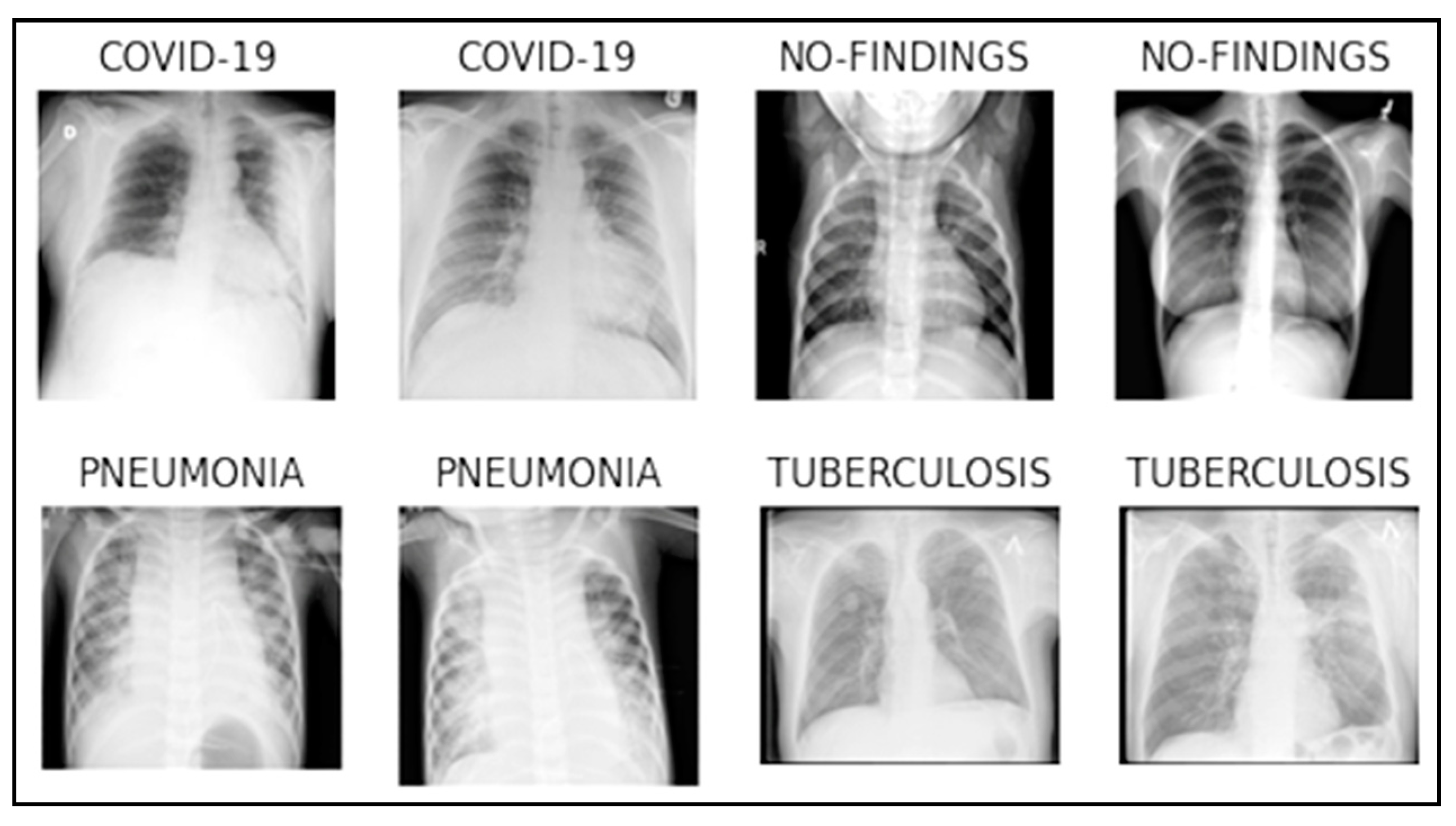
Conditions It Can Detect:
- Pneumonia
- Tuberculosis
- Lung cancer
- Pleurisy
- Emphysema, bronchitis
Advantages:
- Fast and widely available
- Affordable and painless
- Detects pneumonia in early stages
- Performed in nearly all clinics
Preparation:
- No special preparation required
- Takes only 5–10 minutes